

Category Archives: Uncategorized
Hip Strength Predicts Non-Contact ACL Injury
As discussed in previous postings, there is growing consensus in the sports medicine community that hip muscle weakness is contributory to various knee injuries. For those involved in high-level competitive sports, the ACL (anterior cruciate ligament) is the most frequently ruptured ligament of the knee joint. Although risk factors for ACL tears are multifactorial, a new prospective study published by our group in the American Journal of Sports Medicine reports that an athlete’s hip strength predicts future non-contact ACL injury. Non-contact ACL tears result from an athlete’s inherent movement patterns (as opposed to direct blow or contact from an external object or player) during high-velocity multidirectional sports (such as basketball, soccer, football, and volleyball).
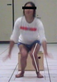
How exactly does hip strength influence the likelihood of ACL injuries? Basically, impaired hip strength is thought to underlie specific movement patterns that place excessive stress and strain on the ligaments and tendons of the knee. As evident in the figure below, there is significant inward collapse at the knee upon landing from a jump. Weakness and/or lack of control of the hip muscles (i.e. gluteus medius and gluteus maximus) is thought to be contributory to this inward motion of the knee. Interestingly, this specific movement pattern also has been shown to be a predictor of ACL tears in a previously published prospective study.1
So to what degree does hip strength influence ACL injury risk? To answer this question, we followed 501 competitive athletes (138 females and 363 males) over the course of a single sports season. Athletes deemed high risk for injury based on their preseason hip strength had their probability of ACL tears more than double. On the other hand, athletes classified as low risk based on their preseason hip strength test had their probability of ACL tears decrease by nearly 90%. These findings applied equally to men and women.
What does this mean for athletes concerned with tearing their ACL? Impaired hip strength more than doubles your risk of suffering an ACL tear. This is why at MPI we examine hip strength as part of our Return to Sport Testing Protocol. Also, we place a high emphasis on hip strength and movement retraining as part of our ACL rehabilitation and injury prevention programs. Regardless of where you do your training or rehabilitation however, you should consider dedicating more time to hip strengthening—your knees will thank you for it!
References
- Hewett TE, Myer GD, Ford KR, et al. Biomechanical measures of neuromuscular control and valgus loading of the knee predict anterior cruciate ligament injury risk in female athletes: a prospective study. Am J Sports Med. 33:492-501, 2005
This post was co-written by Rachel K. Straub, MS, CSCS
Improving Shock Absorption after ACL Reconstruction: Protecting Against Future Arthritis?
ACL injury is a common sports injury. It has been well documented that persons who have undergone ACL reconstruction have a higher risk of developing knee osteoarthritis. It is thought that poor recovery of hip and knee muscle strength and/or poor movement mechanics post ACL-reconstruction, may in part, contribute to the accelerated progression of knee osteoarthritis in this population.
The body has 2 types of shock absorbing systems: the passive system and active system. The passive shock absorbers are the viscoelastic properties of cartilage and bone, while the active shock absorbers are the eccentric action of muscles. In theory, the more one uses the active shock absorbers (i.e. the hip and knee extensors), the less reliance on the passive shock absorbers (i.e. stress on bone and cartilage of the knee joint).
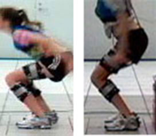
In a recent paper published in the American Journal of Sports Medicine, we demonstrated that improving active shock absorption by increasing hip and knee flexion during a single leg landing task resulted in a decrease in knee joint compressive forces. On average, knee compressive forces decreased by 61% of bodyweight (81 lbs) when subjects were instructed to land more softly (i.e. bend more at the hip and knee) when jumping off a 10 inch platform. These findings suggest that teaching patients to land with greater hip and knee flexion decreases knee joint loading and may prevent or delay the development of knee arthritis in persons who have undergone ACL reconstruction.
Evaluation of shock absorption capability is an important part of the Return to Sport Test at MPI. Using high speed cameras, we evaluate a patient’s ability for active shock absorption during sport specific tasks (i.e. jumping/landing and cutting). In turn, teaching patients to improve active shock absorption is a critical component of our Return to Sport Training program. Using force plates and cameras, we can provide instant feedback to patients regarding their landing performance thereby facilitating the process of motor learning (i.e. muscle memory). By improving use of the hip and knee muscles during sport specific tasks, we seek to minimize the potential for knee pain and arthritis following ACL reconstruction (see example below).
Hip Strength & Control: The Key to Protecting the Knee
After 20 years of studying the mechanics of knee injury and publishing over 100 scientific papers on this topic, I have come to one simple conclusion: most knee injuries stem from poor hip strength and/or control. While this may sound like a fairly strong statement, in reality, it makes a lot of sense. Consider the following line of reasoning:
1) The knee joint is the articulation (connection) of the femur (thigh bone) and tibia (shin bone).
2) As such, the femur comprises half the knee joint.
3) Femur motion is controlled by the hip musculature; particularly the gluteus maximus and gluteus medius.
4) Therefore, the hip muscles control half of the knee joint!
In a recent publication, I discuss the influence of altered hip mechanics on 3 of the most common knee conditions seen in orthopaedic practice: patellofemoral pain, iliotibial band syndrome and anterior cruciate ligament injury. In this paper, I review the growing body of scientific literature suggesting that strength and motion impairments at the hip, pelvis and trunk may contribute to knee injuries. Interestingly, research suggests that females are more likely to exhibit these tendencies than males.
Based on the recent evidence being published in clinical, biomechanics, and sport medicine journals, interventions that address impairments proximal to the knee may be beneficial for patients who present with various knee conditions. More specifically, a biomechanical argument can be made for the incorporation of pelvis and trunk stability as well as dynamic hip joint control into the design of knee rehabilitation programs. While I am not discounting the importance of quadriceps and hamstring strength with respect to knee mechanics, I argue that there should be EQUAL emphasis on both hip and knee strength. Unfortunately most rehabilitation protocols (particularly for patellofemoral pain and ACL reconstruction) do not place enough importance on high level hip muscle strengthening and neuromuscular retraining.
In support of this premise, recent research out of Brazil suggests that hip strengthening may be more important than quadriceps strengthening for the treatment of patellofemoral pain.1-3 Furthermore, a randomized controlled trial published from our group demonstrated that isolated muscle hip strengthening improved pain and functional outcomes in females with chronic patellofemoral joint symptoms.
Obviously, more research needs to be conducted to fully validate the role of altered hip strength and mechanics on knee injury. However the research that is emerging is leading to a paradigm shift in our thinking of knee injury mechanics, rehabilitation and injury prevention strategies. Keep in mind that while the pain may be at the knee, the cause of the symptoms may be coming from somewhere else. Obviously, a thorough biomechanical analysis would be necessary to make this determination.
References
- Fukada TY, Rossetto FM, Magalhaes E, Bryk FF, Lucareli PR, de Almeida Aparecida Carvalho N. Short term effects of hip abductor and lateral rotator strengthening in females with patellofemoral pain syndrome: A randomized controlled clinical trial. J Orthop Sports Phys Ther. 2010;40:736-742.
- Fukada TY, Melo WP, Zaffalon BM, Marconded FR, Magalhaes E, Bryk FF, Martin RL. Hip Posterolateral musculature strengthening in sedentary women with patellofemoral pain syndrome: A randomized controlled clinical trial with 1-year follow-up. J Orthop Sports Phys Ther. 2012;42:823-830.
- Nakagawa TH, Muniz TB, Baldon RM, Maciel D, de Menezes Reiff RB, Serrao FV, Clin Rehab. The effect of additional strengthening of hip abductor and lateral rotator muscles in patellofemoral pain syndrome: A randomized controlled pilot study. 2009;22:1051-1060.